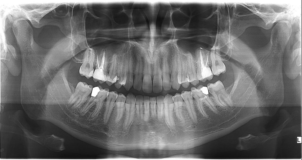
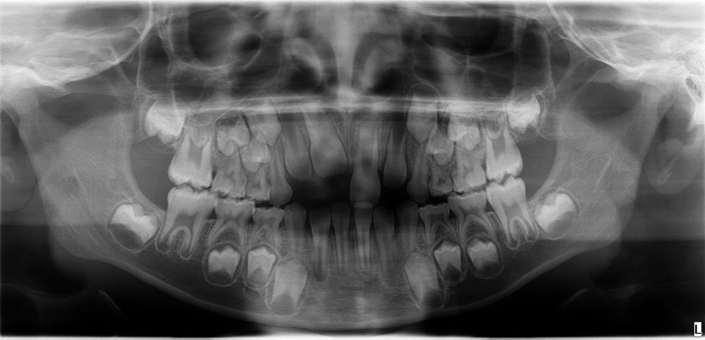
Orthopan or ortopanthomogram
It is the most widely-used dental and facial bone imaging in 2D in stomatology. Orthopan is used to display the upper and lower jaw or all teeth, maxillary sinus, and jaw joints. The image shows the jaw’s bone structure, enabling the study of roots and the changes around them. Orthopan shows where the caries processes are hiding and enables a general inspection of the upper and lower jaw – all in a single image.
The latest technology of the digital Orthopan guarantees a high-quality image that is optimally adjusted with software.
Orthopan imaging is painless and only lasts 16 seconds.
 op300_pan_P1-1024×544
 OP300_P2_Standard-pan-pediatric